


Spatial vision and medical imaging
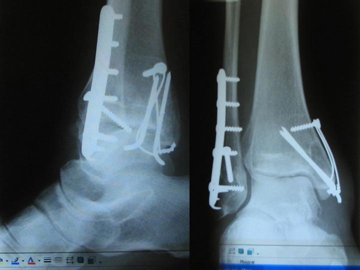
Metals in ankle bones before removing. Image: HUNAGI Visual Resources
 Spatial vision and advanced imaging tools are inevitable in medicine.
Out of the scopes of the geospatial information, but there are analogues: medical imaging provides several hundreds of million imageries per year used for visual interpretation. Present generation of technologies offers 3D and 4D imaging but visual interpretation "spatial thinking" remains a must for the physicians.
Because bones and metals absorbs (attenuate) more of the X-ray passing through them than do the other tissues, they are clearly visible on an X-Ray film. That is why X-ray imageries and particularly those which are taken from two directions enabling 3D visual interpretation are commonly used e.g. in the planning of surgery operation of removing metals. The radiology technology was founded by the discovery of X-rays by Prof Röntgen in 1895, while the first tracer work was done by the Hungarian Georg von Hevesy in 1911.
The radiography today includes mammography, bone mineral densiometry, angiography, cardiatic catheterization. A well established different approach, the ultrasound technology produces today 3D imagery even in case of rapid diagnostic requirement. Recently, 4D ultrasound with temporal resolution opens new vistas in medical applications.
Computers with advanced communications enabled the introduction of teleradiology and computer assisted telediagnostics based on signal and document transmission.
Computers as enabling technology made possible a wide variety of medical imaging technologies including CT (computer assisted tomography the first imaging technology to allow 3D images of the internal structures within a body using by scanned X-ray images processed and visualized as virtual slices, cross section of the body and organs), MRI (magnetic resonance imaging to image chemical processes in the body), MRS (magnetic resonance sprectroscopy, measuring concentrations of metabolites and imaging chemical processes), SPECT (single-photon emission computed tomogrpahy) , and PET (positron emission tomography, which creates images by detecting subatomic particles). Common feature of these advanced tools are that high capacity computers connected with imaging hardware are using to reconstruct sectional or other images from complex data sets, while the interpretation of the visualised results is allowed real-time during the ongoing data acquisition.
Source: http://dsp-psd.communication.gc.ca/Collection/H118-13-2003E.pdf
More on advanced medical imaging: 3D Imaging in Medicine 2nd Ed., CRC Press, 2000ISBN: 084933179X). The book surveys clinical applications of 3D imaging currently pursued, including detailed information on computer integrated surgery, radiation therapy, CT angiography, orthopedics and motion analysis of joints, illustrates methods using software readily available on the Internet and provides over 100 color images that effectively portray details, particularly for 3D renditions.
Co-author of book is Gabor T Herman of the City University of New York, a close institutional partner of the Department of Image Processing and Computer Graphics of the Szeged University of Sciences. The Department lead by Prof. Attila Kuba was created in the Institute of Informatics in 2003. In 2005, Prof. Gabor T.Herman participated a Workshop hosted by the Szeged University of Sciences dedicated to the memory of its mathematician László Kalmár (1905-1976) (http://www.inf.u-szeged.hu/kalmar2005/) . The curricula of the Department contains also the spatial informatics as lectured subject. Note, Szeged University of Sciences is member of HUNAGI via the Department of Physical Geography and Geoinformatics headed by Prof. László Mezősi.
posted by RFG @ 5/29/2006 10:26:00 AM
![]()
![]()


0 Comments:
Post a Comment
<< Home